The conjunctiva is the clear, thin membrane that covers the white of the eye. Years of UV light, wind, and dust can thicken small clusters of that tissue. Over time, this recruits new blood vessels and slowly pushes the growth onto the cornea. Once the growth crosses onto the cornea, it becomes a pterygium, and it can distort the corneal curvature with surprising precision. The long-established surgical treatment is conjunctival autograft. During this procedure, Dr. Bradley Barnett removes the abnormal tissue and covers the exposed area with a small patch of the patient’s own healthy conjunctiva.
Decades of peer-reviewed evidence support this approach, and in real-world review data, conjunctival autograft recurrence rates cluster around 5 to 9 percent, depending on technique and fixation method. The technique works, and the science is well established. Suture-based autograft is no longer the only durable option, and at California LASIK & Eye in Sacramento, we do not default to yesterday's approach when a better one is available.
Dr. Barnett performs pterygium excision paired with Biovance 3L Ocular, a triple-layer decellularized human amniotic basement membrane graft supplied through Defeye for patients in Sacramento, Roseville, Folsom, Elk Grove, and Davis. During the procedure, the membrane is placed over the surgical bed without sutures or fibrin glue, using a technique documented in peer-reviewed ophthalmology literature.
- What Is a Pterygium (Surfer's Eye)?
- What Are the Symptoms?
- Do I Actually Need Surgery?
- How Does Pterygium Removal Work at California LASIK & Eye?
- Why Biovance 3L Changes the Recurrence Conversation
- What Is Recovery Like?
- What Does Self-Pay Pterygium Surgery Cost?
- Who Is a Good Candidate?
- Why Patients Choose Dr. Bradley Barnett
- Patient Stories and Results
- Frequently Asked Questions
- Book Your Pterygium Consultation
What Is a Pterygium (Surfer's Eye)?
A pterygium is a benign, wedge-shaped growth of conjunctival tissue that extends onto the cornea, most often on the nasal side of the eye. It develops slowly, over years, in response to chronic UV light, wind, dust, and ocular surface dryness. Long-term UV exposure is the most consistent environmental risk factor identified in ophthalmic epidemiology. The nickname surfer's eye reflects the pattern seen in outdoor athletes, farmers, construction workers, and anyone who spends significant time in bright, dry, or windy environments. In the Sacramento Valley, that describes a large share of the population.

Do I Actually Need Surgery?
Not every pterygium requires surgery. Small, asymptomatic growths are typically monitored with UV-protective sunglasses, lubricating eye drops, and periodic exams. The American Academy of Ophthalmology recommends surgical removal when a pterygium threatens vision, causes persistent irritation, or affects appearance significantly. At California LASIK & Eye, Dr. Barnett only recommends surgery when the clinical picture supports it. We recommend only what is right for each patient, even if that is watchful waiting.
Why Biovance 3L Changes the Recurrence Conversation
Recurrence is the single most important outcome in pterygium surgery. Historic bare-sclera excision techniques carried recurrence rates as high as 40 percent or more. Modern conjunctival autograft lowers that to roughly 5 to 9 percent, and fibrin-glue fixation trends toward the low end of that range, closer to 4 to 6 percent in long-term follow-up data. Biovance 3L Ocular is a triple-layer, decellularized human amniotic basement membrane. The decellularization process removes pro-inflammatory chorion and residual donor cellular debris while preserving extracellular matrix proteins such as collagen, laminin, and fibronectin. In a 2026 retrospective review published in Ophthalmology and Therapy, a glueless, sutureless pterygium excision technique using Biovance 3L Ocular reported no recurrences and no pyogenic granuloma, dellen, or infection events in in the study cohort, an encouraging early-stage finding, though longer-term, larger studies are still accumulating. For patients, that translates into three practical advantages: shorter surgical time, no sutures irritating the eye during healing, and a graft designed to lower the risk of the pterygium returning.
What Is Recovery Like?
Most patients return to desk work within 1 to 3 days. The eye is typically red and slightly irritated for 7 to 14 days, and the cosmetic appearance continues to improve over several weeks as new epithelium migrates across the treated area. Contact lens wear is paused for at least 2 weeks, and full healing extends over roughly 4 to 6 weeks. Patients receive prescription antibiotic and anti-inflammatory drops, along with preservative-free lubricating tears. Dr. Barnett schedules follow-up visits at day 1, week 1, and month 1, with additional visits as needed based on healing.

Who Is a Good Candidate?

Candidates for pterygium surgery may experience the following:
- A visible growth of conjunctival tissue crossing onto the cornea
- Persistent redness, irritation, or foreign body sensation despite eye drops
- Blurred or fluctuating vision caused by induced astigmatism
- Cosmetic concerns about the appearance of the eye
- A previously removed pterygium that has recurred
Patients with active ocular surface disease, unmanaged dry eye, or specific autoimmune conditions may need preparatory treatment before surgery.
Dr. Barnett is recognized nationally for his work on the ocular surface and coined the peer-reviewed term STODS (Surgical Temporary Ocular Discomfort Syndrome), which shapes how our team prepares every surgical patient.
Why Patients Choose Dr. Bradley Barnett
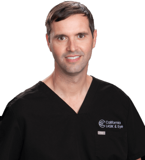
Dr. Bradley Barnett, MD, PhD, is the founder and medical director of California LASIK & Eye. He completed his MD-PhD at Johns Hopkins School of Medicine, his ophthalmology residency at the Johns Hopkins Wilmer Eye Institute, and his fellowship in cornea and refractive surgery at UCSD Shiley Eye Institute. He served as an assistant professor of cornea and refractive surgery at Duke University before founding California LASIK & Eye in 2021. With seven years of post-fellowship surgical experience, Dr. Barnett has performed more than 25,900 procedures, has published 38 peer-reviewed papers including work in Nature Medicine and Science, and holds 5 granted patents in medical device innovation. He is a 2025 Best Cataract Surgeons in America honoree and was the first ophthalmologist in Greater Sacramento to offer several advanced corneal analysis and refractive technologies.
For pterygium patients, three things matter: his cornea fellowship training, his depth of experience with ocular surface disease, and his willingness to say no when surgery is not the right answer. When Dr. Barnett does recommend surgery, patients get a technique built around the latest peer-reviewed evidence rather than the default of a decade ago.
Patient Stories and Results
Book Your Pterygium Consultation
We serve patients across Sacramento, Roseville, Folsom, Elk Grove, and Davis. If a growth on your eye has started to affect your comfort, your vision, or the way you look, contact us to book a consultation with Dr. Barnett to review your options.
Frequently Asked Questions
A pinguecula is a yellowish, raised deposit of protein and fat on the conjunctiva that does not grow onto the cornea. A pterygium is a fleshy conjunctival growth that does extend onto the cornea and can distort vision. Some pterygia begin as pingueculae and progress over time.
Yes. The procedure is performed under topical or local anesthesia, and most patients describe it as comfortable rather than painful. It’s normal to feel pressure or brief sensation during the operation, along with mild irritation for several days afterward.
Most patients drive within 1 to 3 days and return to desk work within a similar window. Outdoor labor, swimming, and contact lens wear are paused for at least 2 weeks. Dr. Barnett gives each patient specific clearance milestones based on their individual healing.
Recurrence is the biggest risk of pterygium surgery. Older bare-sclera techniques carried recurrence rates as high as 40 percent, while modern conjunctival autograft techniques bring that number down to roughly 5 to 9 percent in real-world data, with fibrin-glue fixation trending toward the low end, closer to 4 to 6 percent. Emerging peer-reviewed research on triple-layer decellularized amniotic membrane has reported very low recurrence in early cohorts, though longer-term, larger studies are still accumulating.
Some medical insurance plans cover pterygium excision when the growth threatens vision or causes significant symptoms. Cosmetic-only cases are often not covered. California LASIK & Eye offers transparent self-pay pricing so patients can move forward with surgery without navigating insurance restrictions.
Usually yes, once the corneal surface has healed and astigmatism measurements have stabilized. Removing a pterygium first often improves the accuracy of LASIK, EVO ICL, and cataract lens calculations by restoring a more regular corneal shape.
Consistent UV protection is the most important step. Wear wraparound sunglasses with UV 400 protection, use a wide-brimmed hat outdoors, and treat any underlying dry eye or ocular surface disease. Dr. Barnett builds a prevention plan into each patient’s follow-up care.
- Paganelli B, Sahyoun M, Gabison E. Conjunctival and Limbal Conjunctival Autograft vs. Amniotic Membrane Graft in Primary Pterygium Surgery: A 30-Year Comprehensive Review. Ophthalmology and Therapy. 2023;12(3):1501-1517. https://pubmed.ncbi.nlm.nih.gov/36961661/ Supports: real-world autograft recurrence rates (~5–9 percent), historical bare-sclera comparison, general amniotic membrane recurrence data.
- Li W, Lou Y, Wang B. Recurrence rate with inferior conjunctival autograft transplantation compared with superior conjunctival autograft transplantation in pterygium surgery: a meta-analysis. BMC Ophthalmology. 2021;21(1):131. https://pubmed.ncbi.nlm.nih.gov/33750344/ Supports: autograft technique comparison and general recurrence context.
- Daponte PL, Cigna A, Lescano O, et al. Conjunctival Autograft With Fibrin Glue for Pterygium: A Long Term Recurrence Assessment. Med Hypothesis Discov Innov Ophthalmol J. 2019;8(4):272-277. https://pubmed.ncbi.nlm.nih.gov/31788488/ Supports: fibrin glue autograft long-term recurrence data (single-center retrospective, ~4.4% recurrence). Frame as supporting rather than definitive evidence.
- McDonald M. A Retrospective Review of Glueless, Sutureless Pterygium Excision Using Biovance Triple-Layer Decellularized Amniotic Basement Membrane Tissue. Ophthalmology and Therapy. 2026;15(2):771-780. https://doi.org/10.1007/s40123-026-01311-6 Supports: Biovance 3L Ocular no-recurrence findings and reduced complications in a retrospective cohort. Frame as early-stage evidence pending longer-term prospective data.
- Sliney DH. Geometrical assessment of ocular exposure to environmental UV radiation—implications for ophthalmic epidemiology. Journal of Epidemiology. 1999;9(6 Suppl):S22-32. https://pubmed.ncbi.nlm.nih.gov/10709347/ Supports: UV exposure as the primary environmental risk factor for pterygium.
- American Academy of Ophthalmology. What Is a Pinguecula and a Pterygium (Surfer's Eye)? EyeSmart, reviewed by Cesar A. Briceno, MD. Published January 2026. https://www.aao.org/eye-health/diseases/pinguecula-pterygium Supports: patient-facing definitions, symptom list, standard treatment framework, prevention recommendations.
- Verséa Ophthalmics / DefEYE. Biovance 3L Ocular product specifications, decellularization method, and indications. https://www.versea.com/divisions/ophthalmics/biovance-3l-ocular/ Supports: triple-layer decellularized amniotic basement membrane composition and indications for pterygium repair.